
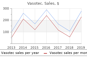
"Buy vasotec 5 mg cheap, hypertension lisinopril".
By: B. Wenzel, M.B. B.A.O., M.B.B.Ch., Ph.D.
Clinical Director, Duke University School of Medicine
Three types of artifacts are visible in this parastenal short axis at the mid ventricular level in a 50-yr-old patient with a left ventricular assist device blood pressure medication in liquid form cheap vasotec 5 mg line. Another type of artifact originates from the fact that ultrasound beams can be wider than the scanline representation on the image hypertension kidney and dialysis specialists generic 10mg vasotec visa. Thus, an ultrasound beam may reflect from a structure slightly off the true axis of the beam, causing a loss in lateral resolution. Mirror image artifacts are frequently seen in the aorta on transesphageal echocardiography. The Doppler principle states that the frequency of a sound (or any wave) will shift (higher or lower) when it is emitted from, or reflected off, a moving object. This occurs because sound waves emitted from a moving source (or reflected off a moving source) are either compressed or expanded depending on the direction of the movement. This is the same principle responsible for the changing frequency of an ambulance siren as it travels toward or away from an observer. In diagnostic ultrasonography, waves are emitted from the transducer at a particular frequency and reflected off moving red blood cells within the heart or blood vessels. If the flow of blood is moving toward the transducer, the sound waves will be compressed (and the frequency of the returning ultrasound will be slightly higher than the emitted ultrasound). The difference between the emitted frequency and the returning frequency is called the Doppler shift. Because the ultrasound machine emits sound at a particular known frequency, the difference between the original ultrasound frequency and the returning ultrasound frequency can be easily determined. This difference in frequency is directly related to the velocity of the structures reflecting the sound (the red blood cells) and, therefore, is related to the velocity of blood flow. This relationship is described by the following equation: v= c(Fs - Ft) 2 Ft (C os) Chapter 1 / Doppler Echocardiography 9. In this case, a reflective structure, such as an area of calcification, causes an "internal" reverberation. The additional "back-and-forth" trip causes the machinery to place an artifactual distal to the original image, but spaced a multiple away from the original distance between the transducer and the reflective structure. Mirror artifacts are commonly seen in the aorta on transesphageal echocardiography as shown in these still frame images. The Doppler principle: when the sound emitting source (in the illustration, the ambulance), is moving toward the listener, the wavelength of the sound waves shorten (or the frequency increases); when the sound emitting source is moving away from the listener, the sound waves will lengthen (the frequency will decrease). Ultrasound emitted from the transducer bounces off moving red blood cells (Bottom), and returns to the transducer. The difference between the emitted frequency and the returning frequency is the Doppler shift. A cardinal principle of digital sampling in general states that the sampling rate must always be at least double the frequency of the waveform being sampled. For example, because humans can hear sounds up to 20,000 Hz, compact discs are recorded using a sampling rate of 44. In Doppler ultrasound, we are sampling the frequency, not of the ultrasound itself, but of the Doppler shift, i. This frequency, as previously discussed, is directly related to the velocity of blood flow. The point at which a waveform cannot be sampled unambiguously happens at a sampling rate of twice the highest frequency that needs to be sampled. Understanding aliasing: aliasing can best be understand by this simple example from sampling theory, the so-called "wagonwheel" example, named after the wagon wheel illusion in old western motion pictures. If we were to "sample" the clock every 15 s (four times per minute) by snapping a picture, we would easily be able to "capture" the motion of the clock, we would see that the hand is rotating clockwise and would be able to discern the rate of rotation. We could tell the rate of rotation, but would not be able to discern the direction. Finally, in the bottom panel, if the velocity of revolution increased to three revolutions per minute (still in the clockwise direction), with the same sampling rate, the perceived direction, based on the sampling, would be counterclockwise, and the perceived rate of rotation would be one revolution per minute.
Persisting eustachian valve in adults: relation to patent foramen ovale and cerebrovascular events blood pressure medication hold parameters purchase 10 mg vasotec overnight delivery. Diagnostic value of transesophageal compared with transthoracic echocardiography in infective endocarditis arteriografia buy genuine vasotec online. Infective endocarditis in the elderly in the era of transesophageal echocardiography: clinical features and prognosis compared with younger patients. He is referred to the emergency department and admitted for rate control, treatment of congestive heart failure, anticoagulation, and consideration of cardioversion. One week before presentation, he noted the onset of increasing fatigue and occasional palpitations when walking briskly to his office. He has been able to continue working full-time in his optometric office, but for the past 2 d, has noted new pedal edema. At presentation to his internist, he is found to be in mild respiratory distress with a respiratory rate of 18/min. Blood pressure is 164/86 mmHg bilaterally, heart rate 120 bpm, irregularly irregular. His point of maximal impulse is in the fifth intercostal space, anterior axillary line. The associated loss of the atrial systolic contribution to left ventricular filling leads to reduced ventricular filling and depressed cardiac output with resultant symptoms of dyspnea and fatigue. The most common associated conditions include a history of systemic hypertension or coronary artery disease. Also to be considered are mitral valve disease (especially rheumatic mitral stenosis), pneumonia/ sepsis, clinical or subclinical thyrotoxicosis (especially in the elderly), pericarditis, pulmonary embolism, pharmaceuticals. Information regarding left ventricular systolic function is frequently helpful for guiding the choice of ventricular rate controlling agent with -blockers used for patients with preserved systolic function and digoxin/diltiazem or a combination for patients with depressed left ventricular systolic function. Current data from both the Atrial Fibrillation Follow-up Investigation of Rhythm Management and Rate Control and Rhythm Control studies suggest cardioversion itself does not reduce the need for chronic warfarin to prevent clinical thromboembolism. Unfortunately, both electrical and pharmacological cardioversion may be associated with clinical thromboembolism, most often occurring during the first 10 days following conversion. For patients with hemodynamic instability, emergent direct current cardioversion is often performed. The use of 34 wk of therapeutic warfarin before cardioversion results in an 80% reduction in clinical thromboembolic risk, to approx 1%. Its anatomical configuration-the narrow neck and multiple ridges-may promote thrombus formation in pathological states. Its external surface appears lobulated and forms part of the cardiac silhouette on chest radiographs. Inferiorly, it sits above the short left main coronary artery as it bifurcates into the left anterior descending and left circumflex vessels, and the great cardiac vein, which flows into the coronary sinus. The use of this "conservative" strategy comes at the "cost" of a delay in cardioversion for the vast majority of patients who could otherwise undergo early and safe cardioversion. The pectinate muscles commence distal to its distinct neck or waist that delineates it from the rest of the smooth-walled left atrium. Composite images showing the internal morphology of the left atrial appendage and pectinate muscles on transesphageal echocardiography. The varied appearance of normal left atrial appearances on transesphageal echocardiography. Normal left atrial appendage morphology can be broadly described as unilobe, bilobed, or multilobed. Color flow Doppler and pulsed wave Doppler examination of the normal left atrial appendage. This contributes to the complex quadriphasic pattern seen on pulsed Doppler examination. Table 4 Transesophageal Echocardiographic Findings in Atrial Fibrillation Finding Spontaneous contrast in left atrium or left atrial appendage Left atrial appendage thrombus Significance Spontaneous echo contrast is thought to increase the risk of thromboembolism.
Vasotec 10mg online. Matlab Project :Continuous blood pressure measurement system by face recognition technique.
Additionally heart attack fever order vasotec american express, whole blood is utilized in patients requiring various types of blood components where it is more economical and practical to do one large transfusion from a single donor than components from possibly multiple donors blood pressure average calculator order generic vasotec from india. An example of this can be seen with canine patients who have ingested anticoagulant-type rodenticides who are suffering both from anemia and decreased clotting factors. Finally, whole blood is often used over components simply because it is the only blood product available to the patient at the time (Callan 2006). Blood product backorders occur on some level within every blood bank facility due to low supply and high demand. This is a safe and accepted practice if the patient can handle the increase in vascular volume seen with whole blood administration. Dogs have approximately 90 mL/kg of blood while cats have approximately 70 mL/kg (Feldman and Sink 2006). Due to these changes, the addition of nutrient and preservative solutions is imperative to help increase red cell viability (Callan 2006). Platelets almost immediately begin to lose viability and function when refrigerated, with a 50% decrease occurring within the first 1218 hours. Approximately 72 hours after storage, there are no longer any viable platelets found within the stored unit (Abrams-Ogg 2000). The loss of labile clotting factors is at a much slower rate than that of platelets. These percentages will continue to decline the longer the unit is left under refrigeration. Once the donor unit has been centrifuged and the red cells separated from the plasma, a nutrient solution such as Optisol is added, and the units are stored in the fridge at 16°C. Packed red cell units should be stored in either a specialized blood bank fridge, a designated blood product fridge, or in a lab fridge that has a separate area only for the placement of blood products. The units should be stored away from other medications, chemicals, and with space between the units. Red cells are living cells that require enough space for fresh oxygen and carbon dioxide diffusion in and out of the bags. It is recommended that red cell units either be suspended/hung in the refrigerator or laid flat; not allowing the units to lie on top of each other or be put into bundles (Abrams-Ogg 2000). Red cell units should not be placed in refrigerator drawers as oxygen exchange may be inhibited by drawer moisture/air controls. One example of this is the chronic renal failure patient; in which erythropoietin is poorly produced and subsequently, bone marrow production of red cells into the circulation is decreased, leading to anemia. Plasma products require an anticoagulant but do not require the addition of a nutrient solution as there are no living cells within the preparation. The use of a freezer without an automatic defrost cycle is recommended (Abrams-Ogg 2000; Schneider 2006). This includes the treatment of hemophilia A and B, anticoagulant rodenticide toxicities, von Willebrand disease, and other factor deficiencies. SelectEmergency/CriticalCareTopicsandTherapies 511 widely accepted for treatment of coagulopathies and/or hypoproteinemic states (AbramsOgg 2000; Brooks 2006). The supernatant is expressed off, and the small amount of precipitate remaining is the cryoprecipitate. Cryoprecipitate is the treatment of choice for hemophilia A, von Willebrand disease, and hypofibrinogenemia. In recent years, Animal Blood Resources International, a merger of Animal Blood Bank and Midwest Blood Bank, has introduced a lyophilized cryoprecipitate product for both canines and felines. The cryoprecipitate, marketed as part of their HemaGold line, is collected through Cohn cold ethanol fractionation to get a 98% pure cryoprecipitate product. The cryoprecipitate is then lyophilized, or freeze dried, allowing the product to be stored in the refrigerator for up to 12 months and quickly reconstituted at the time of use. The cryoprecipitate may be administered at a dosage of 12 mL/kg with severe deficits requiring up to 5 mL/kg. Lyophilized cryoprecipitate can also be used as a pretreatment prior to surgery if a potential coagulopathy is of concern. The manufacturers do caution that because this product has been derived from canine donor plasma that the same risk of infectious disease applies as with other plasma products (Hale 2009) (Table 21.
Delayed complications can also be seen such as vomiting blood pressure chart poster 5 mg vasotec mastercard, aspiration pneumonia what is pulse pressure yahoo cheap vasotec 5mg on-line, tube removal, tube migration, peritonitis, and stoma infection (Hand et al. Gastrostomy tube placement is the technique of choice of long-term enteral support. These tubes are well tolerated by the patient, produce minimal discomfort, allow feeding of either gruel recovery diets or blenderized commercial foods, and can be easily managed by owners at home (Willard 1992). Patients are able to eat normally with gastrostomy tubes in place and can easily be nutritionally supplemented via the tube until the patient is totally self-feeding. For patients that are difficult to medicate and require long-term medications, many medicines can also be given through the feeding tube. The major disadvantage of gastrostomy tubes is the need for general anesthesia and the risk of peritonitis (Willard 1992). For animals requiring long-term management, the initial latex Pezzer catheter can be replaced with either low-profile silicone tubes or with Foley-type gastrostomy tubes. Both of these types can be placed through the external stoma site without the endoscope. Sedation or anesthesia may be necessary based on the individual patient (Hand et al. For removal, if the tube has been in place 16 weeks or less, the tube may be simply removed. The tube is grasped with the right hand close to the body wall, with the left hand holding the animal over the rib cage. It is also helpful to ensure that the patient has been fasted and that a towel is placed over the tube site to catch any gastric contents that may be removed with the tube. Depending on where the breakage occurs, the remaining tube pieces may need to be endoscopically retrieved. Larger patients can easily pass retained parts; smaller patients may need to have them endoscopically retrieved. Jejunostomy Tubes Jejunostomy feeding is indicated when the upper gastrointestinal tract must be rested or when pancreatic stimulation must be decreased. Jejunal tubes can be placed either surgically or threaded through a gastrostomy tube for transpyloric placement. Standard gastojejunal tubes designed for humans are unreliable in dogs due to frequent reflux of the jejunal portion of the tube back into the stomach. Due to the small diameter of these tubes and the location, liquid enteral diets are recommended. Because the jejunum has minimal storage capacity compared with the stomach, continuous rate infusion using a syringe pump is the preferred method of delivery. It is recommended that the jejunal tube be left in place for 710 days to allow adhesions to form around the tube site and prevent leakage back into the abdomen (Guilford et al. Completely changing the delivery equipment every 24 hours will help prevent bacterial growth within the system. Clogging is a common problem; a syringe pump may help to decrease the incidence as will flushing well with warm water every 4 hours. When removing, the tube may be simply pulled out after the securing sutures are removed. Parenteral Nutrition When enteral nutrition is not an option as with gut failure, when enteral nutrition could exacerbate a disease. Due to expense, difficulty in obtaining parenteral nutrition solutions, and ongoing need by the patient, short-term use is usually not justified (Remillard et al. Parenteral nutrition uses a modified solution with nutrients that can be absorbed by the cells without passing through the gut first. Parenteral solutions can be used alone or as a supplement to enteral feedings when insufficient caloric intake is seen. But the use of this type of diet is not without controversy due to the possible association between hyperlipidemia and pancreatitis. A dedicated central venous catheter is required, and the special nutrient solution must be properly prepared. Intensive monitoring is necessary; thrombophlebitis and sepsis are serious complications if strict aseptic technique is not followed (Tennant 1996). A transitional period is necessary to wean the patient from parenteral to enteral feedings (Donaghue 1989).