
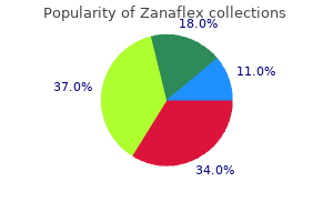
"Purchase online zanaflex, spasms right side of back".
By: S. Mirzo, M.A., M.D., Ph.D.
Vice Chair, Roseman University of Health Sciences
Vitamin B12 is added to fish feeds when necessary in a dry form as part of a multivitamin premix muscle relaxant 5859 order zanaflex american express. Choline has three major metabolic functions: as a component of phosphatidylcholine muscle relaxant review 4mg zanaflex visa, which has structural functions in biological membranes and in tissue lipid utilization; as a precursor of the neurotransmitter acetylcholine; and as a precursor of betaine, which serves as a source of labile methyl groups for methylation reactions such as the formation of methionine from homocysteine and creatine from guanidoacetic acid. Rainbow trout fed a choline-deficient diet developed light yellow-colored livers, protruded eyes, anemia, and extended abdomens (kitamura et al. Lake trout fed a choline-deficient diet for 12 weeks had depressed growth rate and increased liver fat content (Ketola, 1976). Increased liver lipid content has been observed in common carp and channel catfish fed choline-deficient diets (Ogino et al. In addition, common carp developed vacuolization of hepatic cells after being on such a diet for 10 weeks (Ogino et al. A thinning of the intestinal wall muscle and focal degeneration of the exocrine pancreas were observed in choline-deficient sturgeon (Hung, 1989). Channel catfish fed casein-gelatin diets containing excess methionine did not develop signs of choline deficiency; however, catfish fed diets adequate but not excessive in methionine did develop deficiency signs (Wilson and Poe, 1988). Rumsey (1991) has suggested that 50 percent of the choline requirement of rainbow trout can be met from betaine. These observations indicate that certain fish can meet a part of their choline needs through the synthesis of choline by the methylation of ethanolamine, which uses methyl groups from S-adenosyl methionine. Choline is added to fish feeds as a 70 percent choline chloride solution or a 25 to 60 percent dry powder. Choline chloride can decrease the stability of other vitamins in a multivitamin premix during prolonged storage. Inositol is a biologically active cyclohexitol and occurs as a structural component in biological membranes as phosphatidylinositol. Recently, phosphatidylinositol was shown to be involved in signal transduction of several metabolic processes (Mathews and van Holde, 1990). Although similar in many respects to the adenylate cyclase transduction system, the phosphoinositide system is distinctive in that the hormonal stimulus activates a reaction that generates two second messengers. Membrane bound phosphatidylinositol 4,5-bisphosphate is cleaved to release sn-1,2-diacylglycerol and inositol 1,4,5-triphosphate, following the interaction of a hormone or agonist with the receptor on the cell membrane. Inositol 1,4,5-triphosphate stimulates the release of calcium from its intracellular stores in the endoplasmic reticulum, and sn-1,2-diacylglycerol activates protein kinase C to phosphorylate specific target proteins. Signs of inositol deficiency have been reported to include poor appetite, anemia, poor growth, fin erosion, dark skin coloration, slow gastric emptying, and decreased cholinesterase and certain aminotransferase activities in trout (McLaren et al. Rainbow trout fed a diet devoid of inositol had large accumulations of neutral lipids in the liver, increased levels of cholesterol and triglycerides, but decreased amounts of total phospholipid, phosphotidylcholine, phosphotidylethanolamine, and phosphotidylinositol (Holub et al. Inositol appears to be synthesized in common carp intestine (Aoe and Masuda, 1967), but not in amounts sufficient to sustain normal growth of young fish without an exogenous source of this vitamin, because younger carp require a higher level of inositol than older fish. Burtle and Lovell (1989) demonstrated de novo synthesis of inositol in the liver of channel catfish, as well as intestinal synthesis. High concentrations of dietary glucose may increase the need for inositol in some fish (Yone et al. Myoinositol is added to fish feeds when necessary as a dry powder in a multivitamin premix. Ascorbic acid is a strong reducing agent and is readily oxidized to dehydroascorbic acid. Ascorbic acid is a cofactor in the hydroxylation of proline and lysine to hydroxyproline and hydroxylysine in procollagen, which is the precursor of collagen and thus is necessary for the formation of connective tissues, scar tissue in wound repair, and bone matrix (Sandel and Daniel, 1988). Ascorbic acid also facilitates the absorption of iron, thus preventing the anemia often observed in ascorbic acid-deficient fish. In addition, ascorbic acid functions with vitamin E to minimize peroxidation of lipids in fish tissues (Heikkila and Manzino, 1987). Vitamin C-deficient salmon and trout exhibited structural deformities (scoliosis, lordosis, and abnormal support cartilage of the eye, gill, and fins) and internal hemorrhaging usually preceded by nonspecific signs such as anorexia and lethargy (Halver et al. Similar structural deformities such as scoliosis and lordosis due to vitamin C deficiency have been observed in channel catfish (Wilson and Poe, 1973; Andrews and Murai, 1974; Lim and Lovell, 1978; Wilson et al. Japanese eels fed a vitamin C-deficient diet showed reduced growth after 10 weeks and hemorrhage in the head and fins after 14 weeks (Arai et al.
Nutrients muscle relaxant pregnancy discount zanaflex 4mg without prescription, wastes zopiclone muscle relaxant discount zanaflex american express, and hormones are exchanged across the thin walls of capillaries Veins · the actions of muscles to propel blood through the veins Veins carry blood back toward your heart. Interstitial fluid (intercellular fluid) is the fluid found surrounding all the cells of the body Lymph fluid is found in lymph vessels **** these 3 different fluids are the same fluid just with different names depending on where the fluid is located. It also interacts with the blood circulatory system to drain fluid from cells and tissues. The lymphatic system contains immune cells called lymphocytes, which protect the body against antigens (viruses, bacteria, etc. Main functions****** the lymphatic system is composed: lymph vessels lymph nodes organs the functions: to collect and return interstitial fluid, including plasma protein to the blood, and thus help maintain fluid balance to defend the body against disease by producing lymphocytes (B-cells, and T-cells) Lymph organs Lymph organs include the bone marrow, lymph nodes, spleen, and thymus. Lymph Nodes Their two basic functions are: Filtration macrophages destroy microorganisms and debris, bacteria and dead cells are removed from circulatory fluid Immune system activation monitor for antigens and mount an attack against them "X" are lymph nodes throughout the body Spleen Largest lymphoid organ, located on the left side of the abdominal cavity beneath the diaphragm Functions stores disease-fighting components of the immune system (lymphocytes) Immune surveillance and response Cleanses the blood takes out old and defective red blood cells Filtering Lymph nodes are filters of the lymph the spleen is a filter for old red blood cells Disorder of the Lymphatic System Lymphoma is a group of cancers that affect the cells that play a role in the immune system, and primarily represents cells involved in the lymphatic system of the body. They often originate in lymph nodes, presenting as an enlargement of the node (a tumor). Immune System 3 Lines of Defense First line · Skin the skin cannot be penetrated by most organisms unless it already has an opening, such as a nick, scratch, or cut. Second Line · Inflammation is characterized by redness and swelling · Inflammation is stimulated by chemical factors released from damaged cells. The increased blood flow causes puffiness, warmth, and attracts phagocytes (Neutrophils/Macrophages) · Macrophages are giant white blood cells that ingest large numbers of bacteria. Pus is either brained or absorbed by the body Third Line · When the first two systems fail to stop the pathogen the immune system is the last line of defense. Viruses and microorganisms have substances on their outer surfaces that are antigens. Reaction to transplanted organs: the transplanted organ has foreign antigens (proteins from another person). To increase chances for a successful organ transplant, the person receiving the organ should be put on medications to reduce their immune response to the new organ. These drugs will reduce the risk of rejection of the donated organ by the organ transplant patient. Over the next 10-15 days there is a gradual rise in the levels of these products Secondary Immune Response If an antigen enters the body for second time the response is much more rapid. With 1 to 2 days after infection of antigen, high levels of antibodies and immune cells are present in blood. Vaccinations consist of dead or weakened bacteria or viruses that cause an immune response but do not make an individual sick. If the organisms enter the body, the antibodies will be there to attack and kill it. Shot consists of antibodies to the tetanus toxin made in the body of some animal. After 6 months, infant must rely on its immune system to "learn" and acquire immunity to series of diseases. After a pathogen (causes disease or illness) enters the body the person may not have symptoms immediately. It takes time for the pathogen to reproduce & produce enough toxins to make you sick. Antibodies & Antigens · Antigens are "foreign" substances that induce some kind of immune response. Antibody or immunoglobulin is a large Y-shaped protein used by the immune system to identify and neutralize foreign objects like bacteria and viruses. Each antibody recognizes a specific antigen on the surface of the bacteria or virus Vaccinations consist of dead or weakened bacteria or viruses that cause an immune response but do not make an individual sick. Allergy: Immune system produces antibodies /chemicals against a usually harmless substance (antigen) · Allergy is a rapid overreaction to an antigen that is not normally harmful Allergy common symptoms: Runny nose Swollen, ichy eyes Sneezing, coughing A rash these symptoms are causes by the release of histamine which causes an inflammatory response Antihistamines are used to counter these effects Severe allergic reactions can lead to the swelling in the throat which will block the airway. Autoimmune diseases · the immune system fails to recognize some body cells as self. Skeleton muscles generate force and produce movement only by contracting or pulling on body parts. Tendons are attached in such a way that they pull on the bones and make them work like levers. Involuntary Muscles · Involuntary muscles are muscles that are not under your conscious control Involuntary muscles are responsible for activities such as breathing and digesting food Voluntary Muscles Voluntary muscles are under your control, you cause your body to move · Voluntary muscles are used when you smile, turn a page in a book, get out of your chair etc. Types of Muscles · There are three types of muscle tissue skeletal muscle, smooth muscle, and cardiac muscle the skeletal muscles are voluntary muscles the smooth muscle and the cardiac muscle are involuntary 3 types of muscle under microscope Skeletal Muscle · Skeletal muscles help you move (locomotion), they are attached to the bones of your skeleton · At the end of the skeletal muscle is a tendon, which is a strong connective tissue that connects the muscle to the bone.
However muscle relaxant elderly order zanaflex uk, morphological analyses have determined that the spatial patterning of xylem cells occurs temporally prior to the spatial patterning of the phloem cells within the root back spasms 40 weeks pregnant buy zanaflex with american express. Interestingly, vascular defects within the embryonic root have not yet been reported. In the primary root of a wol mutant, there are fewer vascular initial cells, and the entire vascular bundle differentiates as protoxylem. Although this suggests that wol is deficient in procambial, metaxylem vessel and phloem cell specification, a double mutant between wol and fass (which results in supernumerary cell layers) produces phenotypically normal procambial and phloem cells, as well as both protoxylem and metaxylem vessels. Transcriptional master regulators and xylem development Xylem cell differentiation, as marked by secondary cell wall synthesis and deposition, occurs much later in root developmental time relative to protophloem cell differentiation (Figure 11A). However, cells destined to become xylem cells are morphologically identifiable immediately after division of vascular initial cells. In a wol mutant, therefore, there is a lack of cytokinin signaling, a decrease in the asymmetric division of vascular initial cells and ectopic protoxylem cell differentiation in the few remaining vascular cells. This marker then turns on Vascular proliferation-cytokinin signaling Vascular initial cells or stem cells are the progenitor cell type for all vascular cells within the primary root. Regulation of vascular initial cell division is the first step in vascular development and is accomplished, in part, by the two-component cytokinin Insights into Plant Vascular Biology 317 in metaxylem cells and subsequently turns off again prior to secondary cell wall differentiation. This gene is expressed towards the end of the elongation zone in protoxylem cells and then later in metaxylem cells. Together, these findings suggest that, although there are no morphological markers of protoxylem cell specification early in developmental time, there are indeed molecular markers and two distinct developmental states for protoxylem and metaxylem cells: an "early" state and a "late" state. The developmental time point at which the patterning of xylem cells is first regulated by these transcription factors remains unknown. In an apl mutant, protophloem cells are misspecified as protoxylem cells, and there is a short root phenotype (Bonke et al. Pericycle cell specification and differentiation Pericycle cells have been divided into two populations based on gene expression and function. One marker of xylem pole pericycle cell differentiation is the J0121 enhancer trap that marks xylem pole pericycle cells after they exit from the meristematic zone and pass through the elongation zone (Parizot et al. A marker of intervening cells within the pericycle tissue layer helped identify phloem pole pericycle cells. These cells are marked by expression of S17, a basic leucine zipper transcription factor. The function of phloem pole pericycle cells has not been determined, nor are there histological markers of phloem pole pericycle differentiation. However, phloem pole pericycle cells have a very distinct expression pattern and underlying transcriptional signature from that of xylem pole pericycle cells, as determined by expression profiling of marked populations of both of these cells relative to other cell types in the Arabidopsis root (Brady et al. Interestingly, the expression pattern of phloem pole pericycle cells more closely reflects that of cells in the developing phloem cell lineage. No early markers of either phloem or xylem pole pericycle cells have been identified, nor have regulatory factors been Phloem cell patterning and differentiation Only a few factors are known to regulate phloem cell patterning and differentiation, despite protophloem being histologically evident quite early in development relative to xylem cell types (Mahonen et al. In the ops-1 mutant, cell elongation, callose deposition and cell wall thickening fail to occur within the phloem cell lineage (Truernit et al. Finally, in response to external auxin, which is sufficient to induce periclinal divisions in xylem pole pericyle cells, reduced periclinal divisions were observed in the phb-1d mutant. Based on these data, genes were identified whose expression fluctuates over root developmental time;. A rigorous statistical method was developed to identify cases of dynamic expression between roots (Figure 11C), and many of these were expressed specifically in root phloem cell types or in xylem pole pericycle cells. Their function was inferred to be associated with energy capture and lateral root initiation, respectively (Orlando et al. Together, these data indicate that oscillatory, rhythmic and fluctuating gene expression within roots and between roots in the root vasculature serve to regulate patterning of vascular cells, and likely other vascular biological functions. Spatio-temporal regulation of root and shoot vascular development and connectivity Studies on Arabidopsis root mutants defective in cell proliferation and cell differentiation may provide insight into possible common genetic regulatory mechanisms controlling radial vascular development in shoots and leaves. Mutants such as wol show decreased cell proliferation in root procambium and differentiate completely into protoxylem (Mahonen et al. While the spatial and temporal patterns of cell proliferation during root vascular development are becoming clearer, understanding this mechanism in leaves has proved to be more challenging.
Syndromes
- CT scan of the abdomen
- Eliminating waste through urine and feces
- Excessive bleeding after injury or surgery
- Blood in urine
- Thyroid function tests
- Problems breathing
- Hemorrhoids
- Cataracts
- EMG and nerve conduction velocity studies (sometimes done)
The first type consists of primary sensory areas spasms face discount zanaflex uk, which are the first to receive signals from the ascending sensory pathways spasms while peeing buy zanaflex once a day. Instead, I met Haim Sompolinsky, who had just arrived from Israel for a sabbatical year. Haim had previously developed mathematical models of interacting particles in a magnetic field and was now enthusiastically moving on to interacting neurons. I was hooked by this theory of neural networks, so I followed Haim to Jerusalem for post-doctoral training. We applied ideas from statistical physics to understand when artificial neural networks-that is, networks of computational units modeled loosely after neurons-learn not gradually but suddenly, as if with an "aha! When not engaged in lengthy mathematical calculations, I also learned to speak Hebrew and how to make hummus. Many left their office doors open, so you could pop in and ask questions any time. At the other end of the building were computer scientists working in the field of machine learning-a process by which a computer can "learn" from experience rather than being explicitly programmed. Soon I was inventing algorithms that enabled artificial neural networks to learn, and I developed a mathematical theory of a hindbrain neural circuit called the oculomotor integrator. I continued this work after moving to the Massachusetts Institute of Technology as an assistant professor. My theory of the oculomotor integrator was interesting and even plausible, judging from experimental tests by my collaborator David Tank at Princeton. But others were continuing to propose alternative theories, and the field showed no sign of converging on a consensus. My theory assumed the existence of recurrent connections between integrator neurons. In the 1990s, we had both worked at Bell Labs with Winfried Denk, who had since moved to the Max Planck Institute of Biomedical Research in Heidelberg. There Winfried had built an ingenious automated device that could image the face of a block of brain tissue, and then shave off a thin slice to expose a new face. By repeatedly cutting deeper and deeper into the block, the device could acquire a three-dimensional (3D) image of brain tissue. The second type of neocortex consists of secondary sensory areas, so designated because of their heavy interconnections with the primary sensory areas. The third type of cortex consists of motor areas, which are intimately involved with the control of voluntary movement. These cortical areas receive inputs from thalamic nuclei that relay information from the basal telencephalon and the cerebellum, and they send outputs to motor control neurons in the brain stem and spinal cord. For example, because cortical area 4 sends outputs directly to motor neurons in the ventral horn of the spinal cord, it is designated the primary motor cortex, or M1. Manual reconstruction of the wiring diagram would be prohibitively time-consuming. I decided to work on the problem of speeding up image analysis by computer automation. This computational method significantly improved the speed and accuracy of 3D reconstruction of neurons. However, the method still made errors, so it could not completely replace human intelligence. In 2008, we started creating software that would enable humans to work with the machines to reconstruct neural circuits. This eventually turned into the "citizen science" project called EyeWire, which has registered over 150,000 players from 100 countries since its 2012 launch blog. By coloring, they reconstruct the branches of neurons, which are like the "wires" of the brain (Figure A). In 2014, Nature published the first EyeWire-assisted discovery: a new wiring diagram for a neural circuit in the retina. The discovery suggests a new solution to a problem that has eluded neuroscientists for 50 years: How does the retina detect moving visual stimuli? Figure A Seven neurons in a small volume of retina with their dendrites reconstructed from electron microscopic images. It is plain to see that when we speak of the expansion of the cortex in mammalian evolution, what has expanded is the region that lies in between these areas.
Generic zanaflex 2mg with amex. Как правильно выбрать жиросжигатель. Виды жиросжигателей.